Hip Anatomy

The hip is a ball-and-socket joint, made up of two main parts: the femoral head (the “ball”) at the top of the thigh bone and the acetabulum (the “socket”) in the pelvis. Both surfaces are covered with articular cartilage, a smooth tissue that allows the joint to move easily and painlessly. Inside the joint is synovial fluid, which acts as a natural lubricant to reduce friction during movement.

Surrounding the rim of the socket is the labrum, a ring of tough, rubbery cartilage that deepens the hip socket and creates a suction seal to keep the joint stable and the lubricating fluid contained.
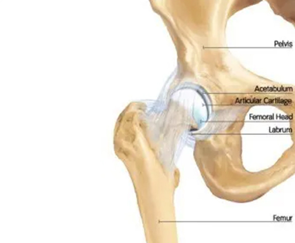
When the labrum is damaged or torn—a common cause of hip pain—this seal is disrupted. As a result, the joint can lose stability and lubrication, leading to increased wear on the cartilage and, over time, the development of arthritis.
The hip joint is also supported by a strong joint capsule, a sleeve of ligaments that helps maintain stability, and a central structure called the ligamentum teres, which connects the femoral head to the acetabulum. Injuries to the capsule or the ligamentum teres can further contribute to hip instability and pain.

Diagnosis of Hip Problems
Diagnosing hip injuries can be complex. Studies show that up to 60% of patients who ultimately require hip arthroscopy were initially misdiagnosed. This is because hip pain can mimic many other conditions, including sciatica, spinal issues, muscle strains, and sports hernias.
Obtaining an accurate diagnosis is critical to ensuring the right treatment. A thorough, systematic approach to diagnosis may include:
- A specialized clinical exam performed by a hip expert
- Targeted X-ray views specifically designed to assess hip anatomy
- Advanced MRI scans performed at centers experienced in pre-arthritic and arthritic hip conditions
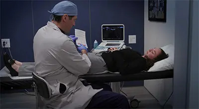
- Ultrasound-guided diagnostic injections into the hip joint to confirm whether pain is originating from within the joint
It’s important to note that many patients remain misdiagnosed even after a physical exam, X-rays, and MRI if those studies are not optimized for early or pre-arthritic hip injuries.