Knee Anatomy
The knee is a complex joint made up of bones, tendons, ligaments, and muscles. All of these structures work together to keep the knee stable and allow smooth movement.
A healthy, well-functioning knee is essential for walking, running, and participating in daily activities or sports. Understanding the knee’s anatomy can help you make informed decisions and discuss the best treatment options with your doctor.
Bones of the Knee
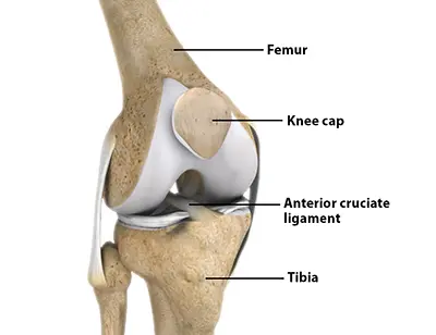
The knee is a hinge joint formed by the thighbone (femur) and the shinbone (tibia). At the end of the femur are two round knobs called femoral condyles, which articulate with the flat surfaces of the tibia called tibial plateaus. The inside plateau is the medial tibial plateau, and the outside is the lateral tibial plateau.
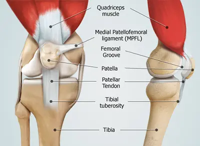
The femoral condyles form a groove on the front of the knee called the patellofemoral groove. The patella, or kneecap, sits in this groove and acts as a shield, protecting the knee from direct trauma.
The fourth bone of the lower leg, the fibula, forms a small joint with the tibia. This joint has very little movement and is not considered part of the main knee joint.
Articular Cartilage and Menisci
All surfaces where bones meet are covered by articular cartilage—a smooth, slippery layer that reduces friction and allows easy movement. This includes the femoral condyles, tibial plateaus, and the back of the patella.
The knee is lined with a synovial membrane that produces synovial fluid. This fluid lubricates the joint and nourishes the cartilage.
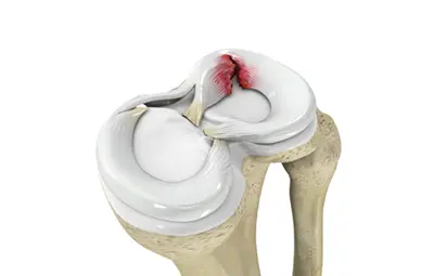
Between the femur and tibia are two C-shaped cartilages called menisci. They act as cushions, absorb shock, and spread the body’s weight evenly across the tibial plateau to protect the cartilage and joint surfaces.
Ligaments of the Knee
Ligaments connect bone to bone and provide stability. The knee has two main types of ligaments:
- Collateral ligaments: On the sides of the knee, they prevent excessive side-to-side movement. The medial collateral ligament (MCL) is on the inside, and the lateral collateral ligament (LCL) is on the outside.
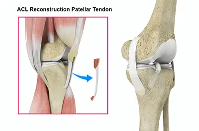
- Cruciate ligaments: Inside the knee, they control forward and backward motion. The anterior cruciate ligament (ACL) is in the front, and the posterior cruciate ligament (PCL) is in the back.
Muscles of the Knee
The quadriceps muscles are at the front of the thigh and straighten the knee when they contract. The hamstrings are at the back of the thigh and bend the knee when they contract.

Tendons of the Knee
Tendons connect muscles to bones. The quadriceps muscles attach to the patella through the quadriceps tendon. The patella then connects to the tibia via the patellar tendon. Together, these structures straighten the knee. The hamstring muscles connect to the knee through the hamstring tendons, enabling bending.