Knee Arthroscopy
What is Knee Arthroscopy?
Knee arthroscopy is a common surgical procedure in which a small viewing instrument called an arthroscope is used to examine, diagnose, or treat problems inside the knee. It is generally a safe procedure, and most patients are discharged from the hospital on the same day of surgery.
Knee Anatomy
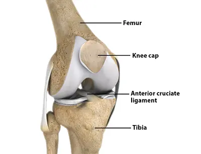
The knee is a complex joint formed where the lower end of the thighbone (femur) meets the upper end of the shinbone (tibia). The patella (kneecap) rests on a groove at the front of the femur, and the fibula, another lower leg bone, forms a small joint with the tibia.
To ensure smooth, pain-free movement, the ends of the bones are covered with slippery articular cartilage. Two C-shaped menisci sit between the femur and tibia, acting as shock absorbers while also providing stability and distributing weight across the joint.
Ligaments, including the cruciate and collateral ligaments, hold the bones together and stabilize the knee. Surrounding muscles attach to the knee bones via tendons, allowing motion. The entire joint is enclosed in a ligamentous capsule lined with a synovial membrane that produces lubricating synovial fluid.
Indications for Knee Arthroscopy
Knee arthroscopy may be recommended for a variety of injuries and conditions, including:
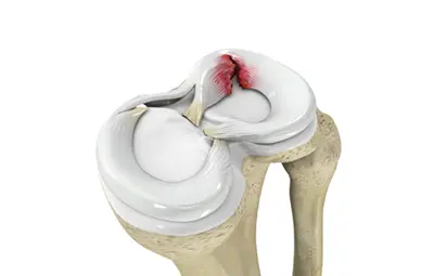
- Torn meniscus
- Torn or damaged cruciate ligaments
- Torn pieces of articular cartilage
- Inflamed synovial tissue
- Misalignment of the patella
- Baker’s cyst, a fluid-filled sac at the back of the knee
- Certain fractures of the knee bones
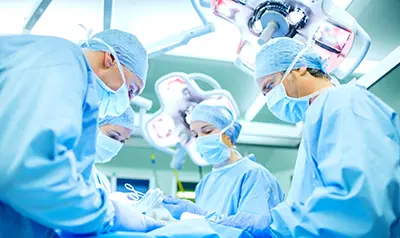
Knee Arthroscopy Procedure
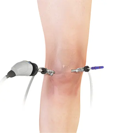
The procedure is performed under local, spinal, or general anesthesia. Two or three small incisions are made around the knee. A sterile saline solution is injected to expand the joint and improve visibility.
An arthroscope, a narrow tube with a tiny camera, is inserted through one incision to allow the surgeon to view the knee on a monitor. Once the problem is assessed, small instruments such as scissors, shavers, or lasers are inserted through other incisions to perform the repair.
Repair Procedures
Repairs may include:
- Removal or repair of a torn meniscus
- Reconstruction or repair of torn cruciate ligaments
- Removal of torn cartilage or loose bone fragments
- Removal of inflamed synovial tissue or Baker’s cyst
- Realignment of the patella
- Microfracture techniques to stimulate cartilage growth
After the repair, the surgeon checks the knee for bleeding or damage, drains the saline solution, closes the incisions with sutures or steri-strips, and applies a sterile dressing.
Postoperative Care Following Knee Arthroscopy
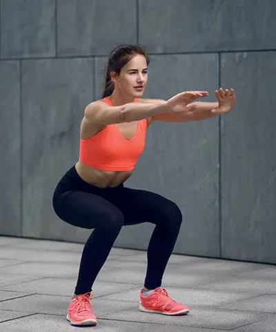
Most patients are discharged the same day. Pain medications are prescribed as needed. Crutches or a knee brace may be recommended for several weeks. A rehabilitation program with therapeutic exercises is usually advised to restore motion and strengthen the muscles around the knee.
Recovery after Knee Arthroscopy
Recovery depends on the type and extent of the procedure. Simple repairs often allow a rapid return to daily activities, and overall recovery from arthroscopy is typically faster than from open knee surgery.