Shoulder Anatomy
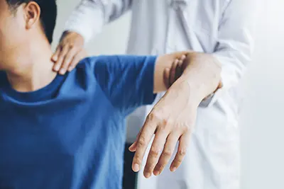
The shoulder is the most flexible joint in the body, allowing a wide range of movements including forward flexion, abduction, adduction, external rotation, internal rotation, and 360-degree circumduction. Despite this mobility, the shoulder is considered the most unstable joint in the body. Stability is provided by the coordinated support of ligaments, muscles, and tendons, which work together to maintain joint integrity while allowing extensive movement.
Bones of the Shoulder
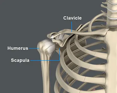
The shoulder joint is a ball-and-socket joint made up of three bones: the humerus, scapula, and clavicle.
Humerus
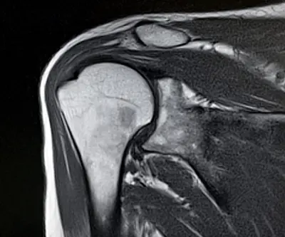
The end of the humerus, or upper arm bone, forms the ball of the shoulder joint. An irregular shallow cavity in the scapula called the glenoid cavity forms the socket for the head of the humerus to fit in. Together, these bones form the glenohumeral joint, which is the main joint of the shoulder.
Scapula and Clavicle
The scapula is a flat, triangular-shaped bone that forms the shoulder blade. It serves as the attachment site for most of the muscles that provide movement and stability to the joint. The scapula has four bony processes: acromion, spine, coracoid, and glenoid cavity. The acromion and coracoid process serve as attachment points for ligaments and tendons.
The clavicle, or collarbone, is an S-shaped bone that connects the scapula to the sternum. It forms two joints: the acromioclavicular joint (with the scapula) and the sternoclavicular joint (with the sternum). The clavicle also protects important nerves and blood vessels that pass beneath it from the spine to the arms.
Soft Tissues of the Shoulder
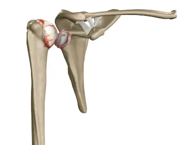
The ends of all articulating bones are covered by articular cartilage, which allows smooth movement and reduces pressure. Extra stability to the glenohumeral joint is provided by the glenoid labrum, a ring of fibrous cartilage that surrounds the glenoid cavity, increasing the depth and surface area to secure the humeral head.
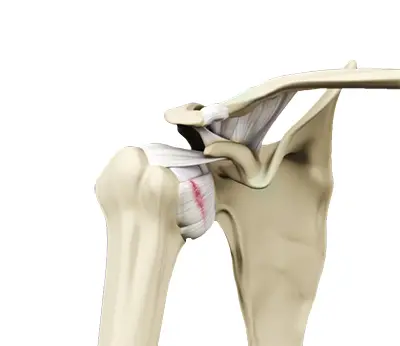
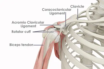
Ligaments of the Shoulder
Ligaments are thick fibers connecting bone to bone. The shoulder ligaments include:
- Coracoclavicular ligaments: These ligaments connect the collarbone to the shoulder blade at the coracoid process.
- Acromioclavicular ligament: This connects the collarbone to the shoulder blade at the acromion process.
- Coracoacromial ligament: It connects the acromion process to the coracoid process.
- Glenohumeral ligaments: A group of 3 ligaments that form a capsule around the shoulder joint and connect the head of the arm bone to the glenoid cavity of the shoulder blade. The capsule forms a watertight sac around the joint. Glenohumeral ligaments play a very important role in providing stability to the otherwise unstable shoulder joint by preventing dislocation.
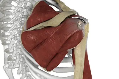
Muscles of the Shoulder
The rotator cuff is the main group of shoulder muscles, forming a sleeve around the humeral head and glenoid cavity. It provides stability while allowing a wide range of motion. The deltoid forms the outer layer of the rotator cuff and is the largest and strongest shoulder muscle.
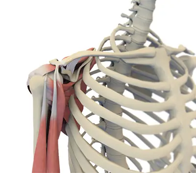
Tendons of the Shoulder
Tendons join muscle to bone, enabling movement. Key shoulder tendons include
- Biceps tendons: long head and short head connecting the biceps muscle to the shoulder
- Rotator cuff tendons: four tendons connecting the humeral head to the rotator cuff muscles, enhancing stability and mobility
Nerves of the Shoulder
Nerves carry motor signals from the brain to muscles and sensory information back to the brain. Nerves pass through the shoulder from the neck, forming the brachial plexus. Main nerves include musculocutaneous, axillary, radial, ulnar, and median nerves.
Blood vessels of the Shoulder
Blood vessels travel alongside nerves to supply the arms. The subclavian artery supplies oxygenated blood to the shoulder, becoming the axillary artery in the armpit and the brachial artery further down the arm. Key veins returning deoxygenated blood to the heart include:
- Axillary vein: drains into the subclavian vein.
- Cephalic vein: in the upper arm, branches at the elbow into the forearm, drains into the axillary vein.
- Basilic vein: runs opposite the cephalic vein near the triceps, drains into the axillary vein.